Biological & Life Science
Manuscript Writing
- New Insights into Complex Role of Mitochondria in Parkinson’s disease
- Antibody Detection in Diagnosis of Schistosomiasis
- Effects of Biomass Particle Size in Cellulosic Biofuel Production
- Dissertation on Modelling Sugarcane Nitrogen Uptake Patterns
- An Integrative Review on Complementary and Alternative Medicine
- A Neuronal Trigger- Endoplasmic Reticulum Stress on Alzheimer’s Disease
A Neuronal Trigger- Endoplasmic Reticulum Stress on Alzheimer’s Disease
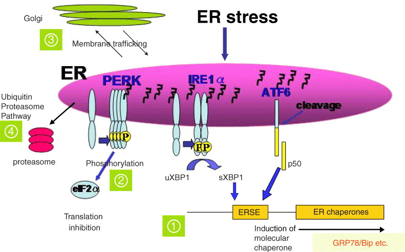
Neurons are vulnerable to different genetic and environmental insults which affect the homeostasis of ER function via the accumulation of unfolded proteins and disturbances in redox and Ca2+ balances. Therefore, it is not surprising that a number of studies have demonstrated that ER stress is present in several neurodegenerative diseases. Evidence of activated UPR signaling has been detected in Alzheimer’s, Parkinson’s and Huntington’s diseases, and in ALS. Furthermore, cerebral ischemia can trigger the UPR, although a concommitant drastic decline in protein synthesis clearly decreases the level of UPR. Viral infections, e.g. Borna virus, can induce prominent ER stress in neurons and subsequently stimulate UPR signaling. In neurons, the tubules and cisternae of ER can extend from the nuclear envelope into dendrites and dendritic spines, and also along axons as far as presynaptic terminals. This means that the neuronal ER is a very specialized organ with functionally different sub-compartments.

Fig.1. Neuronal Trigger by ER Stress in Alzheimer’s Disease
A large body of evidence indicates that the accumulation of intracellular amyloid-β and phosphorylated tau proteins, along with the perturbation of Ca2+ homeostasis, plays a prominent role in the pathogenesis of AD. Interestingly, pPERK staining was abundant in neurons that showed diffuse staining for phosphorylated tau protein, whereas this staining was less prominent in neurons containing neurofibrillary tangles. The tangles themselves were not stained with pPERK. In particular, in hippocampus the CA1, CA4, and subiculum regions contained an abundance of pPERK-positive neurons.
